Rectus Sheath Hematoma Caused by Noncontact Strenuous Exercise
Article information
Abstract
Rectus sheath hematoma (RSH) is an uncommon but well-documented clinical condition. It is usually caused by direct trauma or anticoagulation, although there are many other causes. However, RSH after noncontact strenuous exercise is very rare. We present a rare case of RSH after playing volleyball without direct trauma that was successfully treated by angiographic embolization.
INTRODUCTION
Rectus sheath hematoma (RSH) with inferior epigastric artery injury is an uncommon but well-documented clinical condition [1]. It is usually associated with abdominal trauma or anticoagulation. Other causes of RSH include abdominal surgery, old age, asthma, coughing, and subcutaneous drug injection into the abdomen [2]. Some cases of RSH due to direct trauma or other causes have been reported, but very few for RSH caused due to noncontact strenuous exercise. We present a rare case of RSH after playing volleyball without direct trauma in a healthy individual.
CASE REPORT
A 41-year-old woman with right lower quadrant abdominal pain presented to the emergency department. She had experienced abdominal pain after playing volleyball a week previously, which soon resolved with rest. But, the pain recurred after spiking the ball while playing volleyball on the day of admission. The pain aggravated by strengthening her abdomen and subsided with rest. There was no history of direct abdominal trauma. She had no medical history and was not on any medication. Her vital signs were stable: blood pressure, 110/60 mmHg; pulse rate, 87 beats/min; respiratory rate, 16 breaths/min; and body temperature, 36.2°C. Physical examination revealed tenderness and a palpable mass in the right lower quadrant of the abdomen. Laboratory data, including hemoglobin levels, in the normal range. Computed tomography (CT) revealed a 6×4×18 cm rectus sheath hematoma with extravasation in the right lower rectus abdominis muscle (Fig. 1). In addition, angiography revealed bleeding of the inferior epigastric artery branch (Fig. 2). Angiographic embolization was performed, and extravasation was not observed 3 days later (Fig. 3). The patient was discharged on day 7 without complications. On follow-up CT at 3 months, RSH was completely absorbed (Fig. 4).
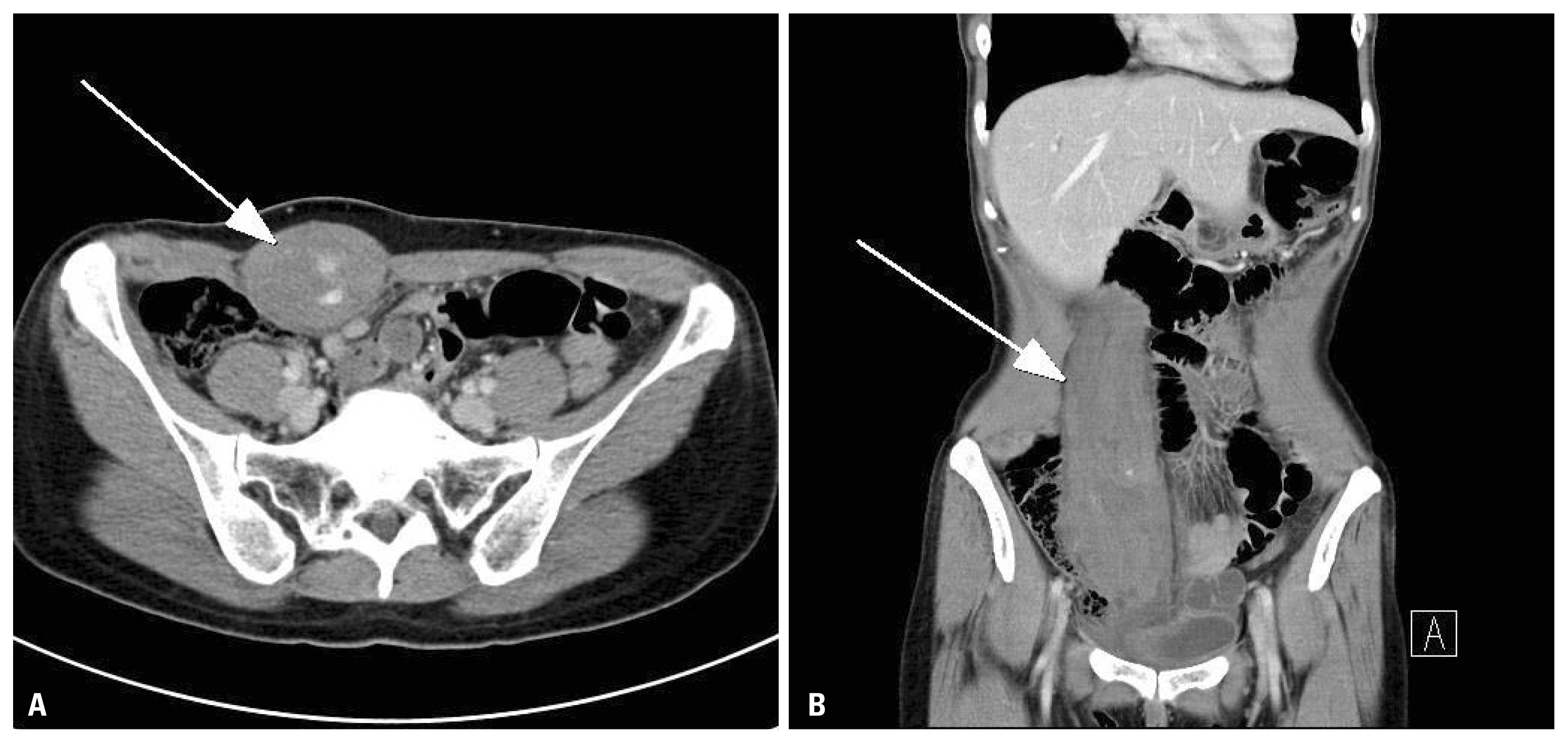
Abdominal CT shows rectus sheath hematoma on (A) right lower quadrant with extravasation (arrow) and (B) on right rectus abdominis muscle (arrow). CT: computed tomography.

Angiography shows extravasation of lower branch of right inferior epigastric artery (arrow).

Extravasation of right lower quadrant was not observed 3 days after angiographic embolization (arrow).

Rectus sheath hematoma was completely absorbed 3 months later.
DISCUSSION
RSH is an uncommon cause of abdominal pain, with an incidence of approximately 1.8%. It usually occurs in women in their fifth or seventh decades [3]. Anatomically, the posterior sheath does not exist below the arcuate line of Douglas. Because there is no protection from the posterior rectus sheath, abdominal pressure is directly applied to the inferior epigastric vessels [2].
Various causes of RSH include abdominal trauma, anticoagulation therapy, abdominal surgery, old age, asthma, coughing, and drug injection into the abdomen. RSH due to twisting or sudden position change without a direct blow, such as that during yoga, tennis, and volleyball have been rarely reported [2].
RSH due to strenuous exercise has been described in tennis players, mainly due to trunk hyperextension with eccentric contraction of the abdominal musculature followed by concentric contraction during trunk flexion. The nondominant rectus abdominis muscle is at a high risk because of its primary involvement in the overhead swinging motion, regardless of the sport [4]. Teske [5] reported that RSH was more common on the right side (60%) and in the lower abdominal quadrants (80%).
RSH was diagnosed based on patient history and physical examination. It is important to distinguish RSH from other abdominal diseases such as appendicitis or diverticulitis. The patient may have a rigid abdomen and tenderness. A palpable mass or bluish discoloration can be found on the injured abdomen. CT is the radiological modality of choice for diagnosing RSH, revealing any extravasation in case of ongoing bleeding from the epigastric vessels [6,7].
Most cases of RSH are usually self-limiting and can be managed conservatively in stable patients [7]. However, blood loss can be massive and even fatal. In cases of RSH with unstable vital signs, extravasation of epigastric vessels observed on CT, expanding hematoma, or ruptured peritoneum, angiographic or surgical intervention may be needed. Surgical management can be performed in an emergency patient with hypovolemic shock. In most cases, identifying the bleeding focus is difficult in the setting of an expanding hematoma. There is a report on proximal ligation of inferior epigastric artery [8]. At present, angiographic embolization is an accepted first line choice for interventional management of RSH [9]. It is very safe and effective, with an overall success rate of 90% and no procedure-related complications [10].
Taken together, although RSH is rare and can be caused by various causes, we should know that noncontact strenuous exercise is one of the causes. Therefore, when a patient presents with abdominal pain after strenuous exercise, clinicians should consider RSH and avoid misdiagnosis.
ACKNOWLEDGEMENTS
This work was supported by clinical research grant from Pusan National University Hospital in 2017.