
Articles
- Page Path
- HOME > J Trauma Inj > Volume 30(4); 2017 > Article
-
Case Report
Traumatic Extrapleural Hematoma Mimicking Hemothorax - Yong Seon Choi, M.D., Soon Jin Kim, M.D., Sang Woo Ryu, Seung Ku Kang
-
Journal of Trauma and Injury 2017;30(4):202-205.
DOI: https://doi.org/10.20408/jti.2017.30.4.202
Published online: December 30, 2017
- 6,852 Views
- 107 Download
- 1 Crossref
Department of Thoracic and Cardiovascular Surgery, Mokpo Hankook Hospital, Mokpo, Korea
- Correspondence to: Soon Jin Kim, M.D., Department of Thoracic and Cardiovascular Surgery, Mokpo Hankook Hospital, 483 Yeongsan-ro, Mokpo 58643, Korea, Tel: +82-61-270-5574, Fax: +82-61-277-0199, E-mail : innocent-blood@hanmail.net
• Received: November 30, 2017 • Revised: December 16, 2017 • Accepted: December 17, 2017
Copyright © 2017 The Korean Society of Trauma
This is an Open Access article distributed under the terms of the Creative Commons Attribution Non-Commercial License (http://creativecommons.org/licenses/by-nc/4.0/) which permits unrestricted noncommercial use, distribution, and reproduction in any medium, provided the original work is properly cited.
ABSTRACT
- After blunt chest injuries, extrapleural hematoma may result in a collection of blood between the parietal pleura and the endothoracic fascia. Extrapleural hematoma is frequently misdiagnosed as hemothorax. Extrapleural fat sign, the inward displacement of strip of extrapleural fat on computed tomography, is typical radiological findings of extrapleural hematoma. We encountered a case of extrapleural hematoma with a presentation similar to hemothorax after blunt chest injury.
- Blunt chest injuries account for more than 15% of injuries in trauma patients [1]. After blunt chest injuries, patients may have chest wall destruction and lung parenchymal injuries. These injuries may cause hemothorax or pneumothorax, the most common complication that can occur immediately after blunt chest trauma [2]. Rarely, chest injury may result in a collection of blood between the parietal pleura and the endothoracic fascia, named extra-pleural hematoma [3]. Extrapleural hematoma is often overlooked on conventional radiologic studies [4]. The authors report a case of traumatic extrapleural hematoma with a presentation similar to hemothorax.
INTRODUCTION
- A 70-year-old male was admitted to the emergency department after experiencing a blunt traumatic injury from a cultivator traffic accident. At the time of admission, his blood pressure was 150/80 mmHg, heart rate was 93 beats/min, respiratory rate was 18 breaths/min, and body temperature was 36.4°C. He had a history of surgery for lung cancer. He complained of severe chest pain and exhibited paradoxical chest movement. Initial laboratory tests revealed a white blood cell count of 12,510/mm3; -hemoglobin, 13.5g/dL; -platelets, 165,000 mm3, and normal arterial blood gases.
- Contrast-enhanced thoracic and abdominal computed tomography (CT) scans showed regional right hemothorax with contrast-media leakage, subcutaneous emphysema and multiple rib fractures (right 2nd–7th) (Fig. 1). The patient was transferred to the operating room to control the chest wall bleeding and reduce thoracic instability.
- Under general anesthesia, the patient was placed in the supine position and double-lumen endotracheal intubation was used for single-lung ventilation. A right anterolateral thoracotomy was made at the fourth intercostal space. The fractured 5th rib was elevated and a moderately sized hematoma was subsequently identified and gently evacuated. The extrapleural space was then exposed and the parietal pleura’s dense adhesions were detected. Bleeding from intercostal artery was controlled with electrocautery. The fractured 4th and 5th ribs were reduced and plate fixation was performed. A 24 French chest tube was inserted through a separate incision.
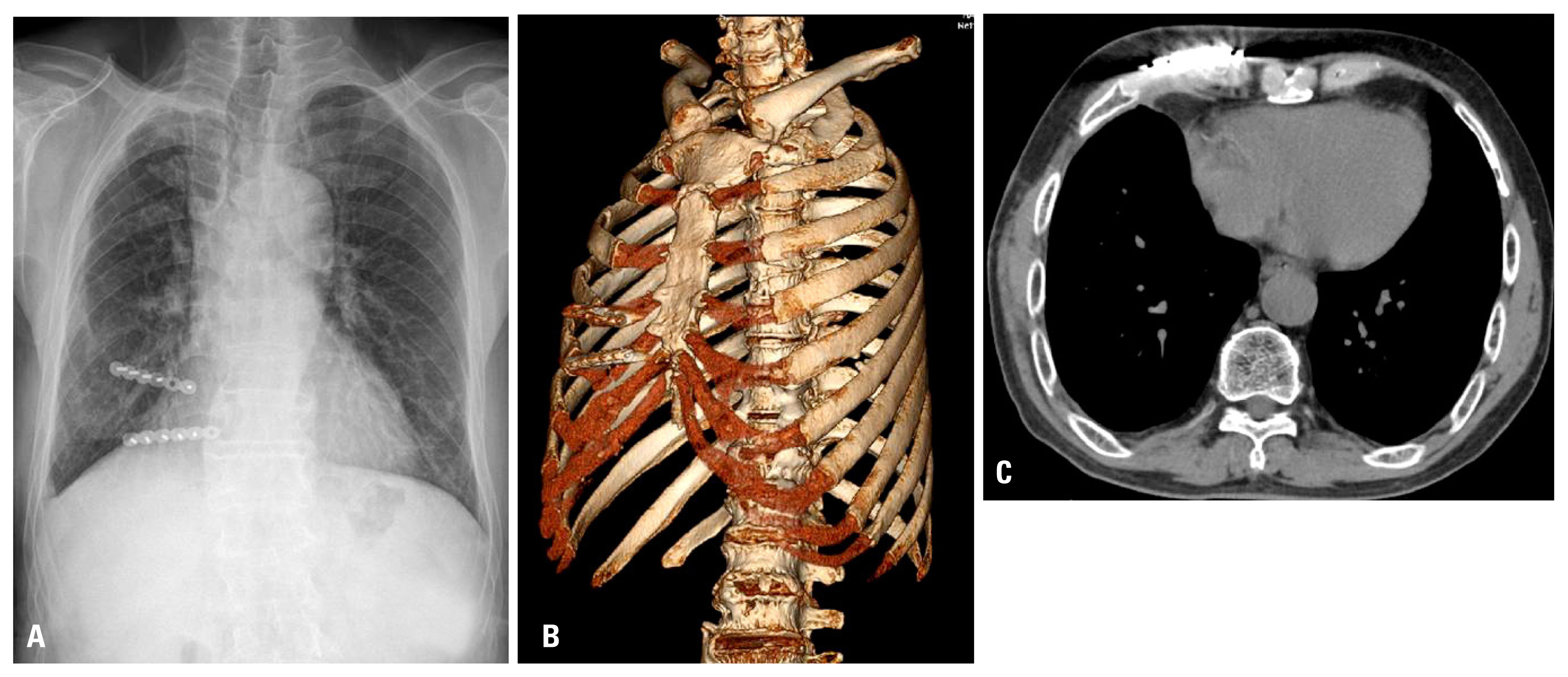
- The patient was extubated 1 day after arrival in the surgical intensive care unit. His postoperative course was uneventful and he was discharged on the 17th postoperative day with no serious complications. The patient is being followed and has experienced no problems for the past 2 months (Fig. 2).
CASE REPORT
- Several cases of hemothorax accompanying blunt chest injury are caused by injury of the chest wall. Most of these cases resolve with conservative treatment such as placement of a drainage catheter [5]. However, intraparenchymal chest tube placement commonly occurs in the presence of pleural adhesions or preexisting pulmonary disease [6].
- The chest wall has a rich vascular system based on the intercostal arteries. After blunt chest injury, the intercostal arteries and veins may tear or rupture. If the parietal pleura remains intact, an extrapleural hematoma may form because the blood cannot escape into the pleural cavity [7]. Similar to our case, patients with thickening of the parietal pleura or with dense adhesions between the parietal pleura and the endothoracic fascia, may ultimately suffer from extrapleural hematoma rather than hemothorax.
- Extrapleural hematoma is frequently misdiagnosed as hemothorax [8]. Typical radiological findings of extrapleural hematoma are biconvex opacity in the involved hemithorax, also known as the displaced “extrapleural fat sign.” The sign is characterized by the inward displacement of strip of extrapleural fat by an extrapleural fluid collection or mass on CT [9].
- In our case, we misdiagnosed the extrapleural hematoma as regional hemothorax, because we overlooked the extrapleural fat sign. Coincidentally, since the contrast-enhanced thoracic CT scan showed contrast-media leakage, we chose surgical intervention rather than tube thoracostomy. The patient’s severe chest pain with dyspnea and paradoxical chest movement also lead us to consider surgical therapy. The choice to operate ultimately enabled us to confirm the presence of a hematoma in the extrapleural cavity.
- We encountered a case of extrapleural hematoma with a presentation similar to hemothorax after blunt chest injury. The presence of the extrapleural fat sign on CT scans is a typical radiological findings of extrapleural hematoma. Traumatic extrapleural hematoma is rare, its diagnosis can be missed as hemothorax and sometimes can be lethal. Treatment should be considered according to the patient’s hemodynamic condition and extent of the extrapleural hematoma. As our case, surgical management can be an effective treatment option in patients with unstable rib fractures.
DISCUSSION
Fig. 1Chest x-ray (A) and computed tomography scan of the patient at admission. 3-dimensional reconstruction (B), pre-enhanced phase (C), arterial phase (D), venous phase (E). Red arrows demonstrating contrast-media leakage.
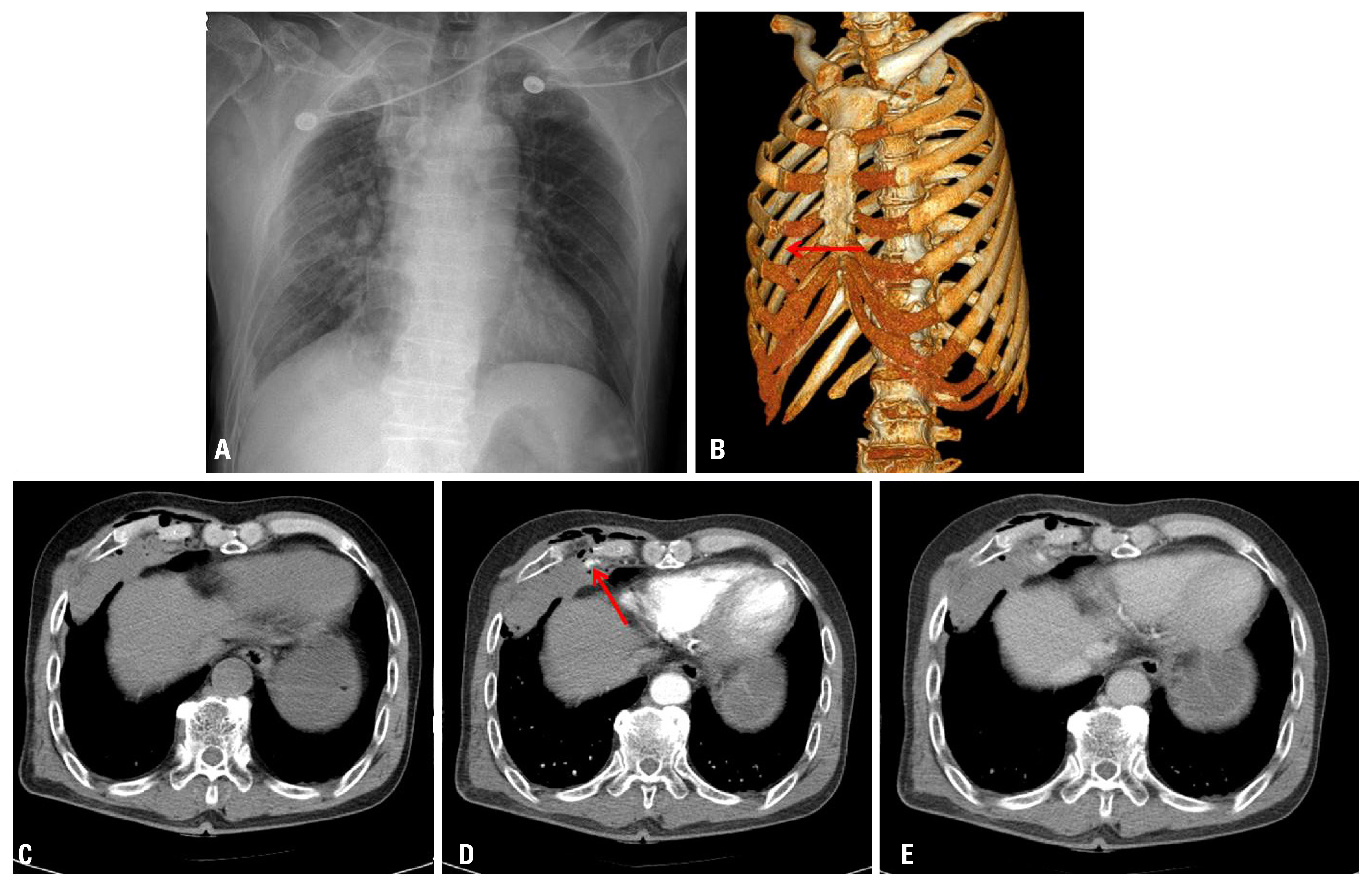
Fig. 2Two month follow-up Chest x-ray (A) and computed tomography scan of the patient. 3-dimensional reconstruction (B), pre-enhanced phase (C).
- 1. Carrillo EH, Richardson JD. Thoracoscopy in the management of hemothorax and retained blood after trauma. Curr Opin Pulm Med 1998;4:243–6.ArticlePubMed
- 2. Ogawa F, Naito M, Iyoda A, Satoh Y. Report of a rare case: occult hemothorax due to blunt trauma without obvious injury to other organs. J Cardiothorac Surg 2013;8:205, ArticlePubMedPMCPDF
- 3. Hii CH, Huong SS, Lo SE, Chiang YH, Tan CK. Extrapleural haematoma: a diagnostic pitfall in blunt chest trauma. Resuscitation 2008;79:348–9.ArticlePubMed
- 4. Santamarina MG, Beddings I, Lermanda Holmgren GV, Opazo Sanchez H, Volpacchio MM. Multidetector CT for evaluation of the extrapleural space. Radiographics 2017;37:1352–70.ArticlePubMed
- 5. Mowery NT, Gunter OL, Collier BR, Diaz JJ Jr, Haut E, Hildreth A, et al. Practice management guidelines for management of hemothorax and occult pneumothorax. J Trauma 2011;70:510–8.ArticlePubMed
- 6. Fraser RS. Lung perforation complicating tube thoracostomy: pathologic description of three cases. Hum Pathol 1988;19:518–23.ArticlePubMed
- 7. Mihos P, Potaris K, Gakidis I, Stathopoulou S. Huge extrapleural hematoma after a blunt chest trauma: an unusual presentation. Eur J Trauma Emerg Surg 2007;33:651–3.ArticlePubMedPDF
- 8. Chung JH, Carr RB, Stern EJ. Extrapleural hematomas: imaging appearance, classification, and clinical significance. J Thorac Imaging 2011;26:218–23.ArticlePubMed
- 9. Aquino SL, Chiles C, Oaks T. Displaced extrapleural fat as revealed by CT scanning: evidence of extrapleural hematoma. AJR Am J Roentgenol 1997;169:687–9.ArticlePubMed
REFERENCES
Figure & Data
References
Citations
Citations to this article as recorded by 
- Hemothorax or not: Use of extrapleural fat sign
Rajat Dahiya, Thaker Nirav, Jaggi Sunila, Talwar Inder
West African Journal of Radiology.2021; 28(1): 18. CrossRef
 PubReader
 ePub Link
 Cite
Cite


